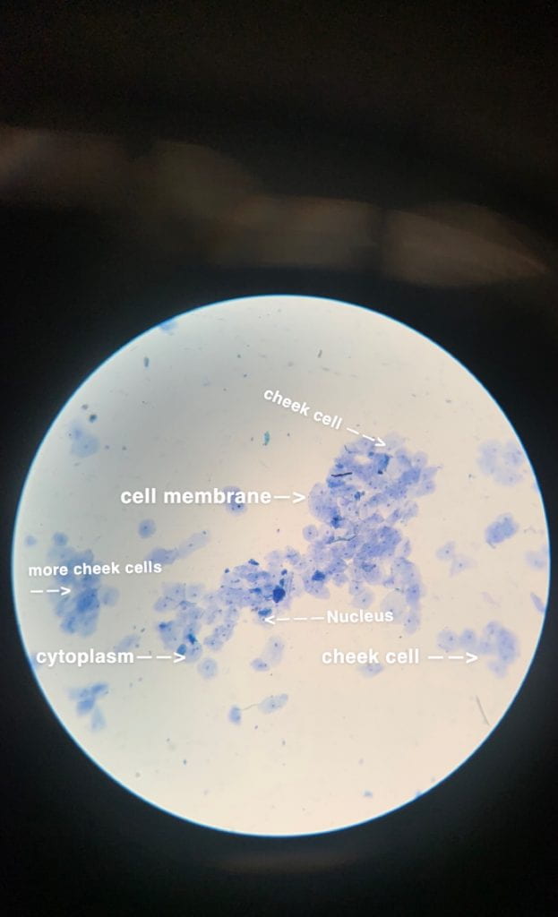
In yesterday’s lab, we took a look at our own cheek cells. In order for this process to happen, we had a few materials like toothpicks, a microscope of course, methylene blue to dye the cells so it will be more visible, and we also learned how to apply a cover slip on the slide. Once we got our materials and everything sorted out, we took a look through the microscope at different powers. First at low power, then medium which was perfect to look through because the nuclei were visible and the cells were crystal clear. When we turned it to high power, the cells as were’t visible anymore so I couldn’t get a picture of them. But besides that, I was able to identify the cell membranes, surrounding the cells, the nuclei which is the small dark blue dots, and the cytoplasm surrounding the nuclei. As you can see, there are other cheek cells scattered around which are known to be dead cheek cells.
Overall, I enjoyed yesterday’s lab. It was an opportunity to get more in depth about microscopes and how to use them. Plus it was a refresher from what I already know about microscopes. I also had fun working with my classmates and my partner, building connections, and discussing what we know about cells as well.